
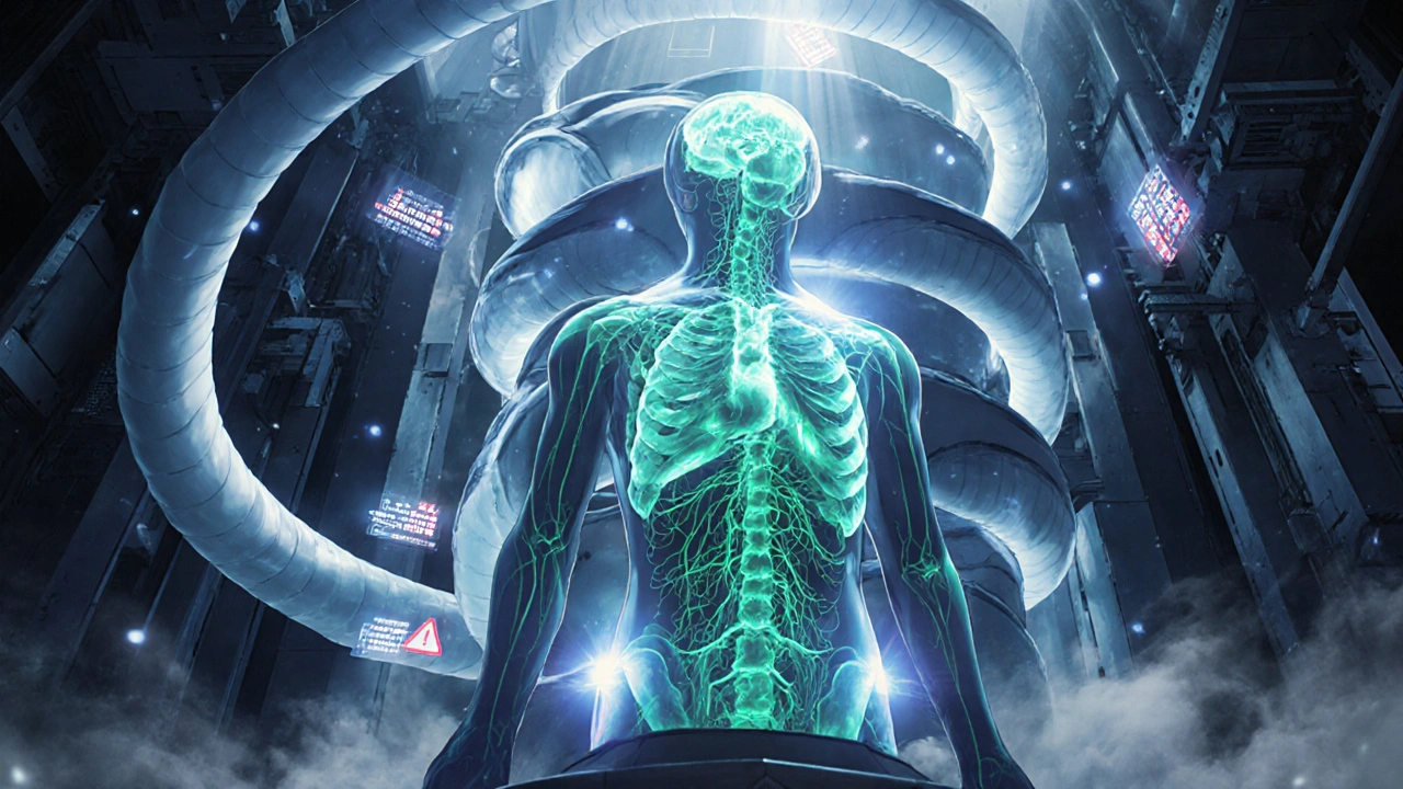
Why accurate cancer staging matters more than you think
Getting cancer staged right isn’t just a technical detail-it changes everything. A misread scan can mean the difference between starting chemotherapy too early, missing a hidden tumor, or delaying surgery that could save a life. That’s why oncologic imaging has become the backbone of modern cancer care. Three tools dominate this space: PET-CT, MRI, and the newer hybrid PET-MRI. Each has strengths, weaknesses, and situations where one clearly outperforms the others. There’s no single best scan for every cancer. The right choice depends on the tumor type, where it’s located, and what question the doctor is trying to answer.
PET-CT: The workhorse of cancer staging
PET-CT became the standard in the early 2000s after its first commercial system launched in 2001. It combines two scans into one: PET shows metabolic activity-where cancer cells are burning sugar at high rates-and CT gives detailed anatomical pictures. Together, they find tumors, see if they’ve spread, and check how well treatment is working. For lung cancer, lymphoma, and colorectal cancer, PET-CT is still the go-to. A 2023 meta-analysis found it correctly identified lymph node spread in non-small cell lung cancer 84% of the time. It’s fast-most scans take 15 to 20 minutes-and widely available. Hospitals from rural clinics to big city centers can run them.
But it has limits. PET-CT uses radiation-about 10 to 25 millisieverts per scan, roughly 500 chest X-rays worth. It also struggles with cancers that don’t use much glucose, like some prostate or kidney tumors. And because CT gives only average soft tissue detail, small tumors in organs like the liver or brain can be missed. That’s why doctors often follow up a PET-CT with another scan if the results are unclear.
MRI: Seeing what other scans can’t
MRI doesn’t use radiation. Instead, it uses strong magnets and radio waves to create incredibly detailed images of soft tissues. That makes it perfect for cancers in the brain, spine, prostate, uterus, liver, and muscles. For prostate cancer, MRI detects tumors with 75% accuracy, far better than older methods. In breast cancer, it finds small lesions hidden in dense tissue that mammograms miss. And when doctors need to tell the difference between scar tissue and returning cancer after radiation, MRI is often the only tool that can help.
Still, MRI isn’t perfect. It’s slow-30 to 60 minutes per scan-and patients have to lie still in a narrow tube. People with metal implants, pacemakers, or certain types of surgical clips can’t have one. It also doesn’t show metabolic activity. A tumor might be visible on MRI, but you can’t tell if it’s alive or dead just by looking. That’s why MRI alone isn’t enough for full staging. It’s best used with other tools.
PET-MRI: The high-end tool for tough cases
PET-MRI, introduced commercially in 2011, merges the metabolic power of PET with the soft-tissue clarity of MRI in a single machine. It’s not common-only about 1 in 5 major cancer centers have one-but when it’s used, the results can be game-changing. For brain tumors, PET-MRI correctly identifies recurrence versus radiation damage 85-90% of the time, compared to just 70-80% for MRI alone. In liver cancer, radiologists report higher confidence in diagnosing metastases with PET-MRI than with PET-CT. A 2023 study in RadioGraphics found that PET-MRI changed treatment plans for nearly half of pancreatic cancer patients because it spotted small tumors others missed.
But there’s a cost. PET-MRI takes longer-up to an hour-and requires more training for technicians. It’s also expensive: $3 to $4.2 million per machine versus $1.8 to $2.5 million for PET-CT. Radiation exposure is cut in half since there’s no CT component, which makes it ideal for young patients or those needing repeated scans. Still, many hospitals hold off because reimbursement is tricky and workflow is complicated. A 2022 study found 63% of sites struggled with PET-MRI’s attenuation correction errors, which can blur images if not fixed by specialized physicists.
How do they stack up? A quick comparison
| Feature | PET-CT | MRI | PET-MRI |
|---|---|---|---|
| Measures metabolism | Yes | No | Yes |
| Measures anatomy | Good | Excellent | Excellent |
| Radiation exposure | High (10-25 mSv) | None | Low (~50% less than PET-CT) |
| Scan time | 15-20 min | 30-60 min | 45-60 min |
| Best for | Lung, lymphoma, colorectal | Brain, prostate, liver, breast | CNS tumors, pelvic cancers, pediatric cases |
| Cost per scan (US, 2023) | $1,600-$2,300 | $1,200-$2,000 | $2,500-$3,500 |
| Availability | Widespread | Widespread | Limited to academic centers |
Which scan for which cancer?
There’s no one-size-fits-all. Here’s what experts use in practice:
- Prostate cancer: MRI is first-line for diagnosis. PSMA PET-CT is added if PSA is rising after treatment. PET-MRI is emerging for high-risk cases where both anatomy and metabolism matter.
- Breast cancer: MRI detects early tumors in dense breasts. PET-CT is used for staging advanced disease. PET-MRI shows promise for monitoring response to chemo, especially in triple-negative subtypes.
- Lung cancer: PET-CT is standard for initial staging. MRI is used if brain metastases are suspected. PET-MRI isn’t routinely used yet but may replace PET-CT in research settings.
- Brain tumors: MRI is essential. PET-MRI is superior for distinguishing recurrence from radiation necrosis-a common diagnostic trap.
- Pancreatic and liver cancers: MRI detects small lesions better than CT. PET-MRI changes management in nearly half of cases, according to recent studies.
- Pediatric cancers: PET-MRI is preferred when possible because it avoids extra radiation. Kids need fewer scans over their lifetime, so reducing exposure matters.

The future: AI and smarter scans
Imaging is getting smarter. In 2024, Siemens Healthineers got FDA clearance for a new PET-MRI system that cuts whole-body scan time to just 6 minutes. At the same time, AI tools are being trained to pull hidden patterns from scans-predicting how a tumor will respond to treatment before it even shrinks. The NCI’s PREDICT trial is testing AI models that combine PET and MRI data to personalize therapy. These aren’t sci-fi-they’re already being tested in hospitals across the U.S. and Europe.
But the biggest shift isn’t hardware. It’s thinking. Doctors no longer ask, “Which scan should we do?” They ask, “What question are we trying to answer?” For a young woman with breast cancer, the goal might be avoiding radiation. For an older patient with lung cancer, speed and availability matter more. The best imaging isn’t the most advanced-it’s the one that answers the right question, at the right time, for the right person.
What’s holding PET-MRI back?
Despite its advantages, PET-MRI hasn’t replaced PET-CT. Why? Three big reasons:
- Cost: The machine is nearly twice as expensive. Maintenance, shielding, and specialized staff add up.
- Workflow: Longer scan times mean fewer patients per day. Scheduling becomes a bottleneck.
- Reimbursement: Insurance companies often pay the same for PET-MRI as for PET-CT, even though the cost is higher. That makes it hard for hospitals to justify the investment.
Still, adoption is growing. About 78% of PET-MRI systems are in academic centers with training programs. That’s where innovation happens. As AI improves image quality and reduces scan times, and as insurers start recognizing the long-term value of fewer repeat scans, PET-MRI will likely become more common-especially for cancers where precision matters most.
What patients should know
If you’re facing cancer imaging, don’t assume one scan is better than another. Ask:
- What’s the goal of this scan-finding the tumor, checking spread, or seeing if treatment is working?
- Is there a reason to avoid radiation, especially if I’m young or need multiple scans?
- Will this scan change how I’m treated? If not, is it really necessary?
Don’t be afraid to ask for a second opinion on imaging choice. The right scan can mean better outcomes. The wrong one can lead to delays, unnecessary treatments, or missed opportunities.


Katie Magnus
November 19, 2025 AT 02:34King Over
November 19, 2025 AT 03:45Johannah Lavin
November 20, 2025 AT 01:20Ravinder Singh
November 20, 2025 AT 23:56Russ Bergeman
November 22, 2025 AT 12:13Dana Oralkhan
November 23, 2025 AT 08:39Jeremy Samuel
November 23, 2025 AT 18:38Destiny Annamaria
November 25, 2025 AT 07:12Ron and Gill Day
November 26, 2025 AT 15:21Alyssa Torres
November 27, 2025 AT 21:31Summer Joy
November 29, 2025 AT 20:00